Since MRE is able to recognize very slight differences in tissue density, there is the potential that it could also be used to detect cancer. This technique is safer and more comfortable for the patient as well as being less expensive than a traditional biopsy. It then pulses sound waves through the liver, which the MRI is able to detect and use to determine the density and health of the liver tissue. The Magnetic Resonance Elastography (MRE) device is placed over the liver of the patient before he enters the MRI machine. NIBIB-funded researchers have developed a method to turn sound waves into images of the liver, which provides a new non-invasive, pain-free approach to find tumors or tissue damaged by liver disease.

Pregnancy-while no effects have been demonstrated on the fetus, it is recommended that MRI scans be avoided as a precaution especially in the first trimester of pregnancy when the fetus’ organs are being formed and contrast agents, if used, could enter the fetal bloodstream.Ĭhronic liver disease and cirrhosis affect more than 5.5 million people in the United States.

Although a causal link has not been established, current guidelines in the United States recommend that dialysis patients should only receive gadolinium agents when essential, and that dialysis should be performed as soon as possible after the scan to remove the agent from the body promptly.

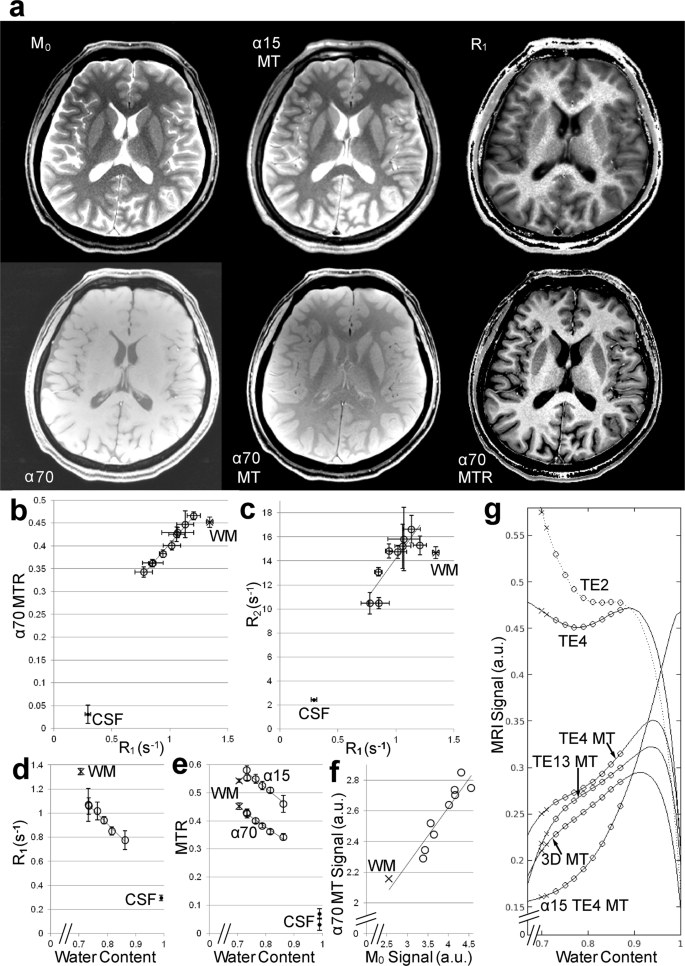
One kind of specialized MRI is functional Magnetic Resonance Imaging (fMRI.) This is used to observe brain structures and determine which areas of the brain “activate” (consume more oxygen) during various cognitive tasks. However, MRI is more expensive than x-ray imaging or CT scanning. Because MRI does not use x-rays or other radiation, it is the imaging modality of choice when frequent imaging is required for diagnosis or therapy, especially in the brain. In the brain, MRI can differentiate between white matter and grey matter and can also be used to diagnose aneurysms and tumors. The brain, spinal cord and nerves, as well as muscles, ligaments, and tendons are seen much more clearly with MRI than with regular x-rays and CT for this reason MRI is often used to image knee and shoulder injuries. They differ from computed tomography (CT), in that they do not use the damaging ionizing radiation of x-rays. MRI scanners are particularly well suited to image the non-bony parts or soft tissues of the body.


 0 kommentar(er)
